
Eyes are living optical systems that bend light, track movement, adjust focus, produce tears, and feed enormous streams of information into the brain. What feels like effortless seeing is actually a nonstop collaboration among transparent tissues, muscles, photoreceptors, nerves, and visual circuits.
The eye is a small organ with a lot packed inside
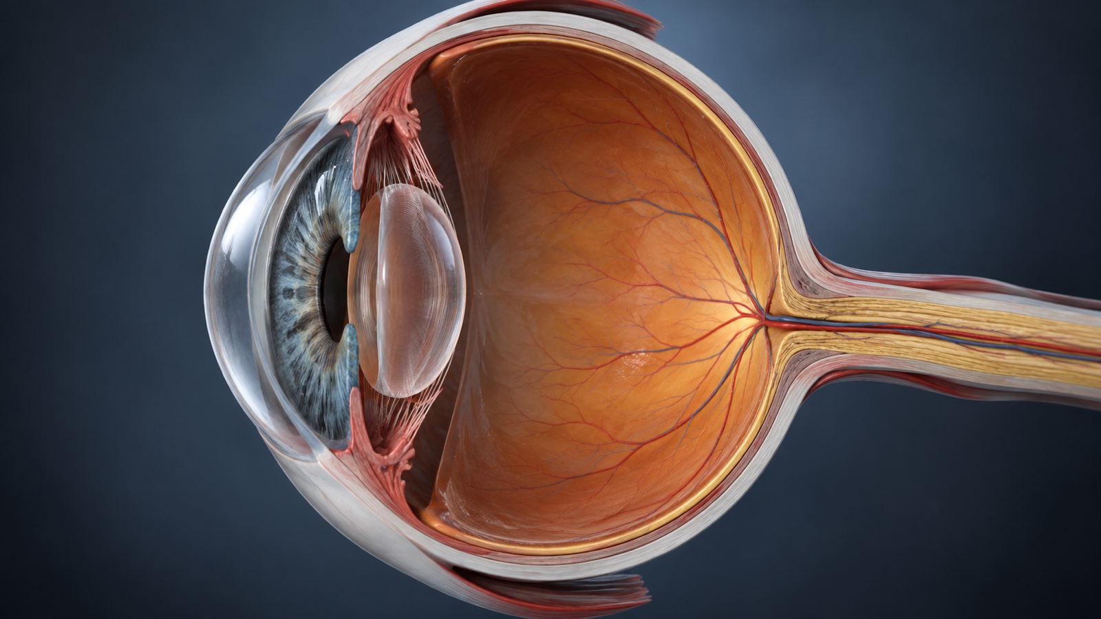
- The adult human eyeball is roughly 1 inch across.
- Most of the eyeball sits protected inside the bony orbit of the skull.
- The visible white part of the eye is called the sclera.
- The sclera is a tough connective-tissue coat that helps maintain eye shape.
- The transparent front window of the eye is the cornea.
- The cornea has no blood vessels in its central healthy state.
- It receives oxygen partly from the air through the tear film.
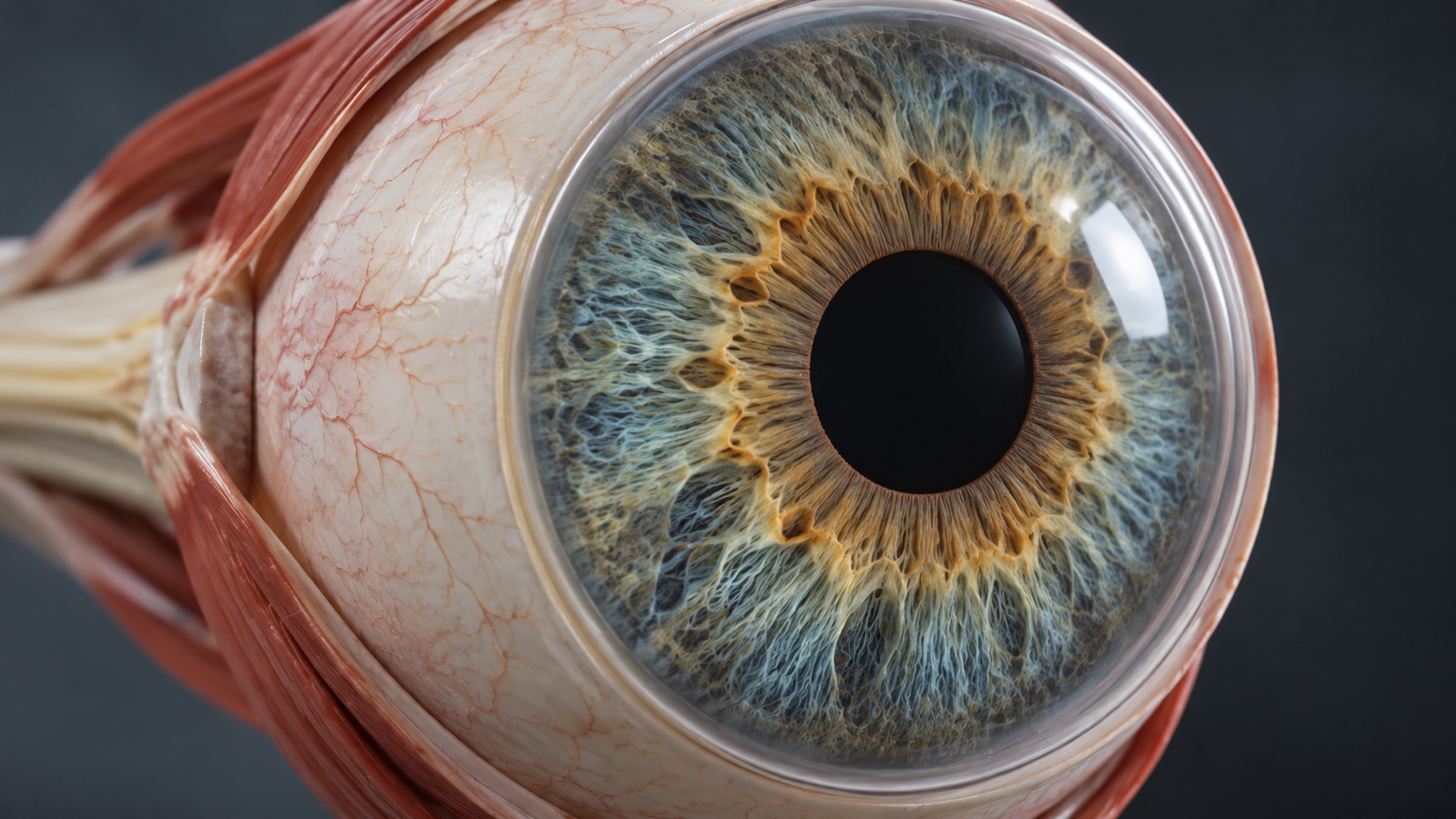
- The colored ring visible through the cornea is the iris.
- The dark opening in the middle of the iris is the pupil.
- The pupil is a hole rather than a black piece of tissue.
- The lens sits behind the iris and pupil.
- The lens is transparent and flexible when young.
- The vitreous humor is a clear gel filling much of the eye behind the lens.
- The aqueous humor is a clear fluid in the front part of the eye.
- Aqueous humor is continually produced and drained.
- Normal drainage helps maintain healthy pressure inside the eye.
- The retina lines the inner back wall of the eye.
- The optic nerve carries visual signals from the retina toward the brain.
- Six main extraocular muscles move each eyeball.
- A structure barely larger than a ping-pong ball manages optics, fluid pressure, motion tracking, light detection, and a direct data cable to the brain all at once.
The cornea and lens bend light into focus
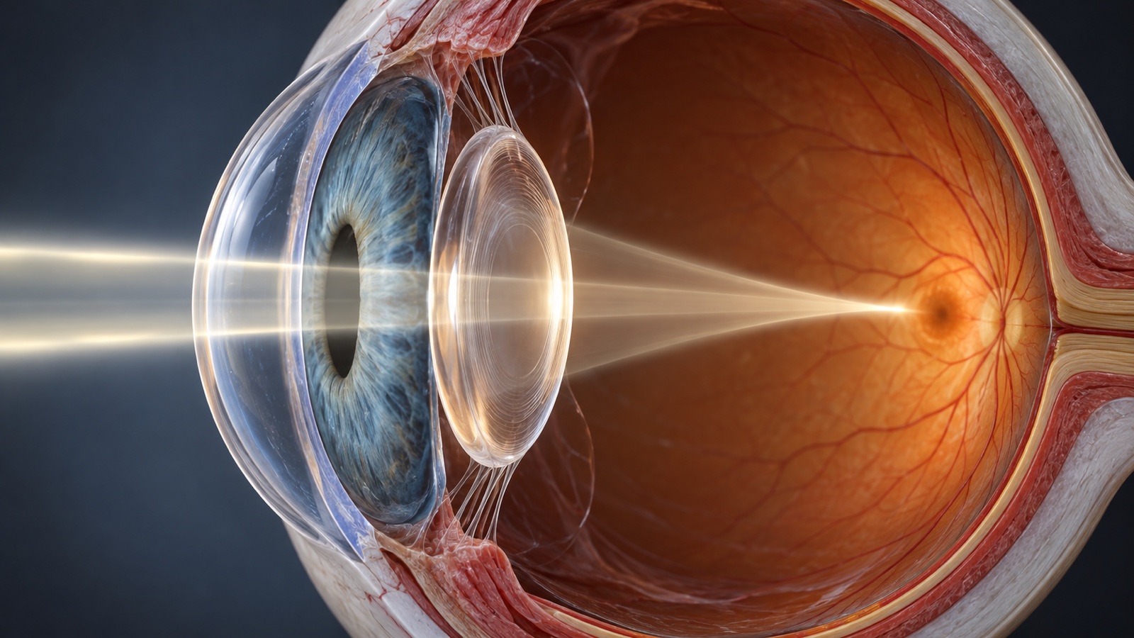
- Most of the eye's focusing power comes from the cornea rather than the lens.
- The cornea bends light because its curved surface separates air from transparent eye tissue.
- The lens fine-tunes focus for objects at different distances.
- Changing lens shape to focus is called accommodation.
- The ciliary muscle helps change the lens shape.
- For near vision, the lens becomes more rounded.
- For distant vision, the lens becomes flatter.
- Accommodation becomes less powerful with age as the lens stiffens.
- This age-related difficulty focusing nearby is called presbyopia.
- Myopia means distant objects focus too far in front of the retina without correction.
- Myopia is commonly called nearsightedness.
- Hyperopia means the optical system tends to focus images behind the retina when accommodation is relaxed.
- Hyperopia is commonly called farsightedness.
- Astigmatism occurs when the cornea or lens has unequal curvature in different directions.
- Glasses and contact lenses redirect light so it focuses more accurately on the retina.
- Refractive surgery changes corneal shape to alter focusing power.
- The image formed on the retina is inverted relative to the outside scene.
- The brain does not need to consciously flip a tiny internal picture like editing software.
- Visual perception develops from neural processing of retinal signals rather than from looking at a literal photograph inside the eye.
- Your eyes are not cameras in the simple sense. They are adjustable biological optics connected to a brain that actively interprets every frame.
The iris controls how much light enters
- The iris contains muscles that change pupil diameter.
- Bright light normally makes pupils constrict.
- Dim light normally makes pupils dilate.
- Pupil changes help regulate how much light reaches the retina.
- Pupils also change size with attention, emotion, medications, and nervous-system activity.
- The two pupils usually respond together when light enters either eye.
- This paired response is called the consensual light reflex.
- The pupil can look perfectly black because much of the entering light is absorbed inside the eye.
- Flash photography can produce red-eye when light reflects from blood-rich tissue at the back of the eye.
- Iris color depends largely on melanin amount and how light scatters through iris tissue.
- Brown eyes generally contain more melanin than blue eyes.
- Blue eye color does not come from blue pigment.
- It arises mainly from light scattering in an iris with relatively little melanin.
- Green and hazel eyes reflect combinations of pigment and scattering effects.
- Eye color is influenced by multiple genes rather than one simple dominant-recessive switch.
- Babies' eye color can change during early life as iris pigmentation develops.
- Heterochromia means the two eyes or parts of one iris differ noticeably in color.
- Heterochromia can be inherited or acquired.
- A sudden new change in iris color can warrant medical evaluation.
- The iris is basically a muscular aperture with genetics, pigment, and optical physics conspiring to make it look decorative.
Rods and cones turn light into neural signals
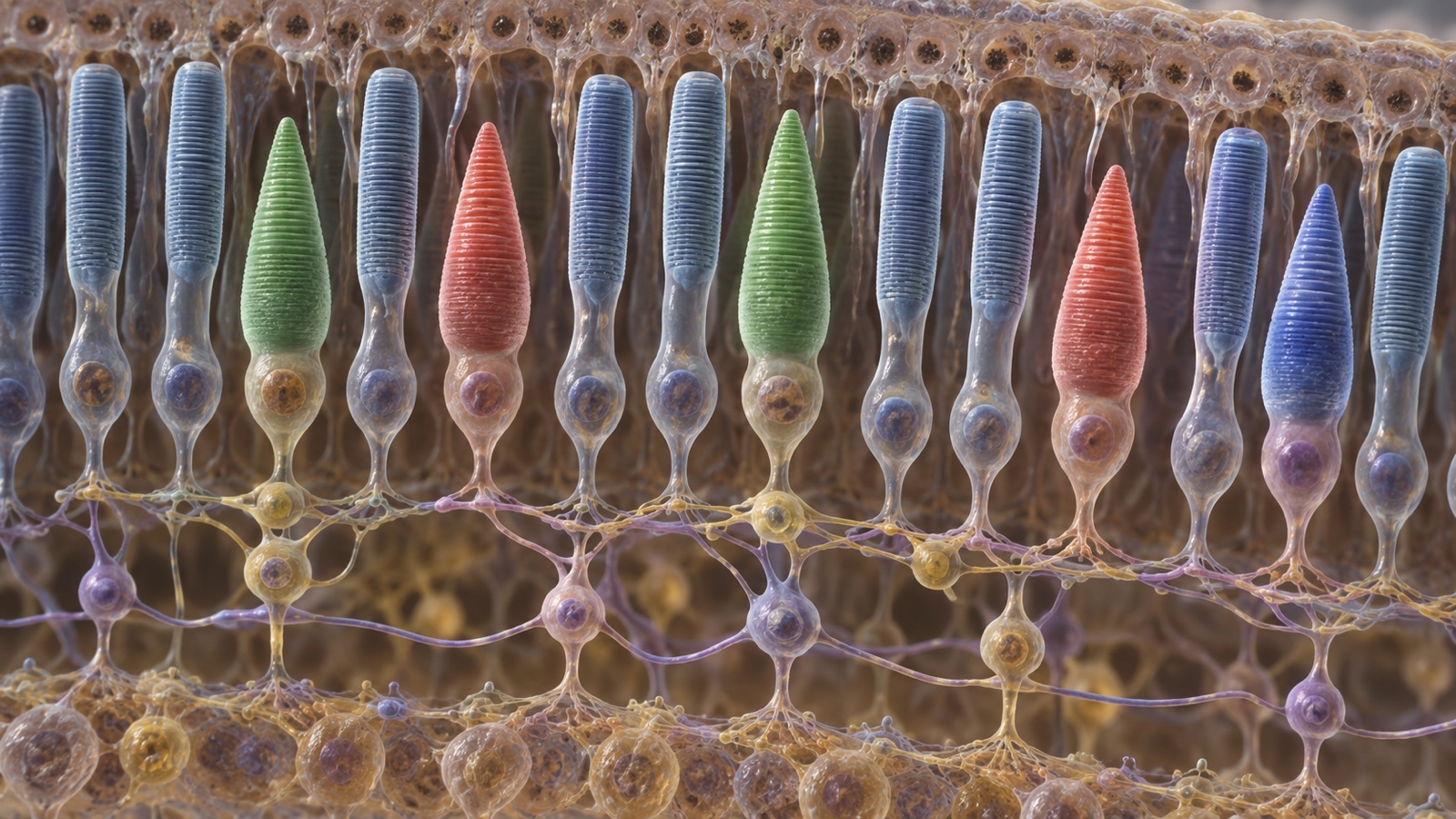
- The retina contains specialized light-sensitive cells called photoreceptors.
- The two main human photoreceptor types are rods and cones.
- Rods are highly sensitive in dim light.
- Rods do not provide normal color vision.
- Cones work best in brighter conditions.
- Humans normally have three classes of cones with different wavelength sensitivities.
- Those cone classes support trichromatic color vision.
- The fovea is a tiny central retinal region specialized for sharp detail.
- The center of the fovea is densely packed with cones.
- Reading small print relies heavily on foveal vision.
- Peripheral retina contains proportionally more rods than the foveal center.
- Peripheral vision is especially useful for detecting movement.
- Photoreceptors convert light into electrical changes through phototransduction.
- Retinal circuits begin processing visual information before signals ever reach the brain.
- Bipolar cells relay information from photoreceptors toward ganglion cells.
- Ganglion-cell axons bundle together to form the optic nerve.
- The optic disc is where the optic nerve exits the retina.
- There are no photoreceptors at the optic disc.
- This creates a natural blind spot in each eye.
- You almost never notice the blind spot because the brain and the other eye smoothly fill in or compensate for the missing information.
Color vision is built by comparing cone signals
- Human color vision depends on comparing activity across different cone types.
- One cone class is most sensitive to longer wavelengths.
- Another is most sensitive to medium wavelengths.
- A third is most sensitive to shorter wavelengths.
- No cone is simply an isolated red, green, or blue pixel.
- Perceived color emerges from combined neural comparisons.
- Color appearance also depends on surrounding colors and lighting.
- The visual system performs color constancy so familiar objects look relatively stable under changing illumination.
- Common inherited color-vision differences affect red-green discrimination.
- Red-green color-vision deficiency is much more common in males because common forms are X-linked.
- Color-vision deficiency rarely means seeing the world only in black and white.
- True complete absence of color perception is rare.
- Some animals see wavelengths humans cannot detect.
- Many birds can detect ultraviolet light.
- Human lenses and corneas block much ultraviolet radiation before it reaches the retina.
- Very bright light can temporarily bleach photopigments and create afterimages.
- Negative afterimages partly reflect adaptation in visual pathways.
- Staring directly at the Sun can damage the retina even when pain is absent.
- The retina itself lacks ordinary pain receptors that would provide a useful warning during solar injury.
- Color feels like a property painted onto objects, but your brain constructs it from overlapping receptor responses, context, adaptation, and comparison.
Tears keep the eye smooth, clear and defended
- A thin tear film coats the front surface of the eye.
- The tear film helps keep the cornea optically smooth.
- Tears provide moisture and nutrients to the corneal surface.
- They also help wash away small particles.
- Tears contain antimicrobial molecules including lysozyme.
- The lacrimal glands produce much of the watery portion of tears.
- Meibomian glands in the eyelids produce oils that slow tear evaporation.
- Goblet cells help produce mucins that allow tears to spread over the surface.
- Blinking redistributes the tear film.
- Humans normally blink many times each minute, although rate changes with activity.
- People often blink less while concentrating on screens or reading.
- Reduced blinking can contribute to dry-eye symptoms.
- Excess tears drain through tiny openings near the inner corners of the eyelids.
- Tears then pass through the nasolacrimal system toward the nose.
- This is why crying can make your nose run.
- Emotional crying uses the same drainage route as ordinary tears.
- Reflex tears increase when the eye is irritated by smoke, onion vapors, or debris.
- Onion cutting releases volatile compounds that irritate eye-surface nerves.
- Contact lenses float on and interact with the tear film rather than sitting directly on bare corneal cells.
- Every blink is a tiny windshield-wiper cycle that restores an optical surface you depend on for nearly every waking second.
Both eyes and the brain cooperate to build depth and stable vision
- Each eye sees the world from a slightly different horizontal position.
- The brain can compare these differences to estimate depth.
- This binocular disparity is especially useful for nearby objects.
- People with one functioning eye can still judge depth using motion, perspective, size, shading, and other monocular cues.
- The eyes make rapid jumps called saccades several times each second during active viewing.
- Saccades move the fovea from one important target to another.
- Smooth-pursuit movements let the eyes follow a moving target.
- The vestibulo-ocular reflex stabilizes gaze when the head moves.
- This reflex moves the eyes in the opposite direction of head motion.
- Without gaze stabilization, the world would smear every time you walked or turned your head.
- The brain suppresses much awareness of visual blur during rapid saccades.
- Eye movements from both sides are usually coordinated.
- Convergence turns both eyes inward for a nearby target.
- Visual signals from the two eyes partially cross at the optic chiasm.
- Information from the left side of visual space is processed mainly in the right cerebral hemisphere.
- Information from the right side of visual space is processed mainly in the left hemisphere.
- The primary visual cortex sits in the occipital lobe at the back of the brain.
- Visual processing continues through multiple pathways involved in motion, shape, color, recognition, and spatial relationships.
- What you consciously see is therefore a processed interpretation rather than an untouched feed from two cameras.
- Your eyes move constantly while perception feels stable because the brain is excellent at hiding how much active correction is happening underneath the experience.
Protection, aging and 5 final eye facts
- Eyelids, eyelashes, eyebrows, tears, blinking, and the bony orbit all help protect the eyes from injury and debris.
- The eye's natural lens becomes less flexible and more yellow with age, affecting near focus and color transmission.
- A cataract is clouding of the eye's natural lens and becomes increasingly common with age.
- Regular eye examinations can detect vision problems and some eye diseases before noticeable symptoms become severe.
- After 145 facts, seeing feels much less passive: every glance depends on transparent tissues, adjustable optics, moving pupils, retinal circuits, synchronized eye muscles, tear chemistry, and a brain that turns electrical signals into a stable visual world.
Quick FAQ
Q: What part of the eye does most of the focusing?
A: The cornea provides most of the eye's fixed focusing power. The lens fine-tunes focus for different distances.
Q: Why do pupils look black?
A: Light entering the pupil is mostly absorbed inside the eye, so relatively little returns to the viewer.
Q: Are blue eyes actually blue?
A: The iris does not contain blue pigment. Blue appearance comes mainly from light scattering in tissue with relatively low melanin.
Q: Why do we have a blind spot?
A: The optic nerve must exit the retina somewhere, and that spot contains no rods or cones.
Q: Can staring at the Sun hurt your eyes even if it does not hurt?
A: Yes. Intense solar radiation can damage the retina without producing a useful pain warning.

Ellie is the owner and sole author of Fun Facts, combining her mechanical engineering background with years of research-driven writing to deliver facts you can trust. Every article is thoroughly fact-checked and routinely updated as new science and sources emerge to keep information accurate and current. Her mission is to make learning delightful while upholding high standards of reliability and transparency.